What is a Mammogram? Do I Need One?

Source: Shutterstock
Females who are 40 years old or above may be advised by their healthcare provider to undergo a mammogram screening every year. Additionally, a mammogram may be recommended if the patient or the doctor notices any potential breast cancer symptoms. This procedure checks for any signs of breast cancer and should not cause excessive worry. Though some people may experience discomfort, most describe it as painless and quick.
What is a mammogram?
A mammogram is an x-ray image of the breast. It can be used for screening or diagnostic purposes. Screening mammograms detect abnormalities in the breast even before any symptoms are clinically present. Breast cancer may not show any physical symptoms in some people until it has progressed significantly. A screening mammogram can detect it early on, making it possible to diagnose and treat the cancer promptly. Diagnostic mammograms are used to investigate reported symptoms and clinical findings or as an additional test to confirm a suspicion from other tests.
Although screening and diagnostic mammograms may use the same type of machine, the latter takes longer and exposes the patient to more radiation. This is because diagnostic mammograms require multiple x-ray images from different angles and magnifications to properly examine and identify suspicious findings, increasing radiation exposure.
How does mammography work?
During a mammogram, a patient’s breast is placed between two specialized plates of a mammography unit which is a low dose x-ray machine specifically designed for taking breast mammograms. The plates compress the breast to flatten it, ensuring high-quality and clear images while reducing radiation exposure. X-rays are produced by an x-ray tube inside the mammography unit and pass through the breast, where they are detected by a flat panel detector on the other side of the breast. Depending on the type of mammography, the image will either be stored in a physical phosphor-coated film or as a digital image.
What do mammograms look like?
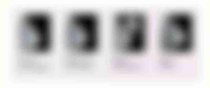
Every woman’s breasts are unique, so mammograms will also vary from person to person, even if there is no cancer present. However, if there are any abnormalities such as cancer, lesions, cysts, or calcifications in the breast, they may show up as unusual white spots or patches on a mammogram.

Normal versus abnormal breast mammograms. Simple benign cysts (second image from the left) tend to have a smooth wall with a regular shape while cancerous tumors often have a more irregular shape (First image from the right). Source: National Cancer Institute
All breasts have dense tissue (fibrograndular tissue) and fatty tissue, which appear differently on a mammogram. Fatty tissue appears as dark areas on a mammogram. In contrast, dense tissue appears white—similar to how breast lesions, cancers, cysts, and calcifications appear on a mammogram.

Varying breast density on mammograms. Source: Densebreast-info.org
Thus, women with dense breasts tend to be more prone to false-negative mammogram results as it can be difficult for radiologists to spot suspicious features in the breast.
Learn more: Breast Density: Are You Informed?
Other types of mammography
Screen-film mammography has been used as a screening tool since the 1970s and has remained the gold standard for breast cancer screening. However, technological improvements have led to the emergence of newer forms of mammography that may and are beginning to replace screen-film mammography in many healthcare institutions.
Digital mammography
Both conventional screen-film mammography and digital mammography use x-rays to produce an image of the breast and follow the same procedures. However, in digital mammography, the x-ray images of the breast are stored digitally instead of on phosphor-coated films in conventional mammography. These digital images can then be digitally manipulated to achieve clearer and more accurate images, which cannot be done in screen-film mammography. In the United States, digital mammography has mostly replaced conventional mammography.
3-D mammography (tomosynthesis mammography)
Also known as digital breast tomosynthesis (DBT), three-dimensional (3-D) mammography uses multiple low-dose x-ray images taken in an arc around the breast, and a computer reconstructs it as a series of thin slices. This is opposed to the x-rays taken from just 2 different angles in conventional and digital mammography. This method has shown to produce a clearer visualisation of the breast tissue and any abnormalities that may be present.
Learn more: New Imaging Technologies and Methods to Detect Breast Cancer
Should I go for a mammogram?
For many, the thought of undergoing a mammogram can be a source of anxiety and discomfort. However, it is important to remember that these tests are essential for the early detection of breast cancer, which can ultimately save lives. Since its widespread implementation, mammograms have contributed significantly to detecting breast cancer early. While some discomfort is expected during the procedure, it is a small price to pay for the benefits of detecting any potential issues early on. If you are unsure whether a mammogram is right for you, it is always best to consult your doctor. They can help you determine if it is appropriate for your situation and provide guidance and support. Ultimately, getting a mammogram is a proactive and responsible choice that can help protect your health in the long run.